
BIOC-200 · Lesson 4 of 12 · 25 min
Protein structure and function
▶ Video coming soon A video version of this lesson is on the way.
A newly made polypeptide is a floppy chain, but within seconds to minutes most chains fold into a precise three-dimensional shape, and that shape is what lets a protein work. An enzyme's active site, a receptor's binding pocket and a channel's pore all depend on side chains being held in exactly the right positions, often to within a tenth of a nanometre. Change the shape and the function is lost: cooking an egg, perming hair and the misfolding that causes some diseases are all changes of protein shape. This lesson describes the four levels at which protein structure is described, the forces that hold a fold together, how proteins fold and unfold, and how two famous proteins, myoglobin and hemoglobin, show structure and function working together.
Four levels of structure
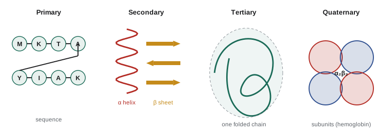
Biochemists describe protein structure at four levels, a scheme proposed by the Danish chemist Kaj Linderstrøm-Lang in the 1950s. The primary structure is the amino acid sequence, together with any disulfide bonds. The secondary structure is the local folding of the backbone into regular patterns held by hydrogen bonds, chiefly the alpha helix and the beta sheet. The tertiary structure is the complete three-dimensional fold of one polypeptide chain. The quaternary structure is the arrangement of several chains, called subunits, in a protein made of more than one chain.
The alpha helix
In 1951 Linus Pauling, Robert Corey and Herman Branson predicted two regular structures that a polypeptide backbone could adopt, working from the planar peptide bond and the best geometry for hydrogen bonds. Both were soon found in real proteins. In the alpha helix the backbone winds into a right-handed coil, and the carbonyl oxygen of each residue forms a hydrogen bond with the N–H group of the residue four places further along the chain. Every backbone C=O and N–H inside the helix is hydrogen-bonded, which makes the helix very stable. The helix rises 0.15 nm per residue and makes a full turn every 3.6 residues, so one turn is 0.54 nm long. The side chains point outward from the coil, like the bristles of a bottle brush.
Some amino acids favour helices and others break them. Alanine, leucine, glutamate and methionine are common in helices. Proline is a helix breaker, because its ring leaves no N–H to donate a hydrogen bond and its rigid shape kinks the chain; glycine is too flexible to hold the helix in place. Many helices are amphipathic: hydrophobic side chains line up along one face, which packs against the core of the protein, while polar side chains on the opposite face meet the water. Every seven residues, about two turns, the pattern repeats, and in coiled coils such as the keratin of hair two amphipathic helices wind around each other with their hydrophobic stripes together.
The beta sheet
In a beta sheet, stretches of nearly fully extended backbone, called beta strands, lie side by side and are linked by hydrogen bonds between the C=O of one strand and the N–H of its neighbour. The strands can run in the same direction (parallel) or in opposite directions (antiparallel), and a sheet may contain both. The side chains point alternately above and below the sheet, so one face can be hydrophobic and the other polar. Because successive residues are 0.33 to 0.35 nm apart along an extended strand, more than twice the rise of a helix, a beta strand of a given length covers much more distance than a helix. Strands are often joined by tight hairpin turns of four residues, which commonly contain glycine and proline.
Silk fibroin is built almost entirely of stacked antiparallel beta sheets, rich in glycine and alanine whose small side chains let the sheets pack tightly. The result is a fibre that is strong because the sheets' backbones are already fully extended and cannot stretch further without breaking covalent bonds, but flexible because the sheets are held to each other only by weak interactions.
Tertiary structure and the forces that hold it
The tertiary structure of a water-soluble protein usually follows one rule: hydrophobic side chains are buried in a core, away from water, and polar and charged side chains are on the surface. The hydrophobic effect described in Lesson 2 is the main driving force for folding. Once the chain has collapsed around its hydrophobic core, many weak interactions lock the fold in place: hydrogen bonds within the backbone and between side chains, ionic interactions or salt bridges between oppositely charged side chains, and van der Waals contacts between atoms packed closely together. The interiors of proteins are packed as densely as crystals of small organic molecules.
Disulfide bonds add covalent cross-links, mainly in proteins secreted into the harsh environment outside the cell. Inside the cell, where the environment is chemically reducing, disulfides are rare. Metal ions can also hold parts of a fold together; the zinc finger, a small structure of about 30 residues found in many DNA-binding proteins, is held together by a zinc ion bound to two cysteines and two histidines.
Each weak interaction is small, a few kilojoules per mole, and folding gives up a great deal of chain entropy. As a result, the net stability of a folded protein is surprisingly small: the free energy of unfolding of a typical protein is only about 20 to 60 kJ/mol, the equivalent of a few hydrogen bonds. Proteins are marginally stable by design. A protein that could never unfold could never be broken down, flexed or regulated.
Folded proteins are built from recurring units. A domain is a part of a chain, typically 50 to 250 residues, that folds independently into a compact structure and often has its own function, such as binding a nucleotide or a calcium ion. Large proteins are often several domains strung together. Within domains, characteristic combinations of secondary structure called motifs or supersecondary structures recur, such as the helix-loop-helix that binds calcium in calmodulin and the beta-alpha-beta unit of many enzymes.
Not all proteins are globular. Fibrous proteins such as collagen, keratin and silk are long and repetitive and provide strength. Collagen, the most abundant protein in the human body, is a triple helix of three chains in which every third residue is glycine, the only residue small enough to fit in the crowded centre, and many prolines are hydroxylated to hydroxyproline. The hydroxylation needs vitamin C, which is why a lack of vitamin C causes scurvy, with weak blood vessels and gums. Some proteins, or parts of them, are intrinsically disordered: they have no stable fold on their own and take up a structure only when they bind a partner.
How proteins fold
In the early 1960s Christian Anfinsen showed that the enzyme ribonuclease could be completely unfolded with urea and a reducing agent, which breaks its four disulfide bonds, and that when both were removed slowly it refolded and regained full activity. The conclusion, for which he shared the 1972 Nobel Prize in Chemistry, is that the amino acid sequence contains all the information needed to specify the folded structure. The folded state is, in most cases, the arrangement of lowest free energy.
Folding cannot happen by a random search. A chain of 100 residues with even three possible conformations per residue has 3¹⁰⁰, about 5 × 10⁴⁷, shapes; sampling one every picosecond would take far longer than the age of the universe, a puzzle known as Levinthal's paradox. Real proteins fold in microseconds to seconds because folding is guided. Local structures such as helices form quickly, the chain collapses around its hydrophobic core, and the protein slides down a funnel-shaped energy landscape toward the native state, with each step lowering the free energy.
In the crowded cell, newly made chains can stick to each other before they fold. Chaperone proteins prevent this. Some, such as Hsp70, bind exposed hydrophobic stretches and release them over and over until the chain folds; others, the chaperonins such as GroEL in bacteria, enclose a single chain in a protected chamber where it can fold alone. Many chaperones are made in greater amounts when cells are heated, which is why they were first named heat-shock proteins.
Predicting a protein's structure from its sequence was one of the great open problems of biochemistry for fifty years. In 2020 the deep-learning program AlphaFold, from DeepMind, predicted structures with accuracy close to that of experiments in a blind test, and its predictions for more than 200 million proteins are now freely available. Demis Hassabis and John Jumper shared the 2024 Nobel Prize in Chemistry for this work with David Baker, who designs new proteins with computational methods. Experimental methods remain essential: X-ray crystallography, nuclear magnetic resonance and cryo-electron microscopy provide the measured structures on which such programs are trained and against which they are checked.
Denaturation and misfolding
Denaturation is the loss of a protein's folded structure without breaking its peptide bonds. Heat denatures proteins by shaking apart the weak interactions; most human proteins unfold somewhere between 40 and 70 °C, which is why a high fever is dangerous and why the white of a fried egg turns solid and opaque. Extremes of pH denature proteins by changing the charges on side chains, which breaks salt bridges and adds repulsion. Detergents such as sodium dodecyl sulfate and chemicals such as urea and guanidinium chloride unfold proteins by making the hydrophobic interior less unwelcome in water. Organisms that live in hot springs make proteins with extra salt bridges and tighter cores that stay folded near 100 °C; the DNA polymerase from one of them, Thermus aquaticus, is the enzyme used in PCR.
Some proteins misfold into a form that sticks to other copies of itself, forming long fibres called amyloid in which beta strands stack across many chains. Amyloid deposits are found in Alzheimer's disease, Parkinson's disease and type 2 diabetes. In prion diseases, such as Creutzfeldt-Jakob disease in people and bovine spongiform encephalopathy in cattle, a misfolded prion protein converts normal copies to the misfolded form, so the disease can be transmitted by a protein alone. Sickle-cell disease shows how one changed residue can matter: replacing glutamate with valine at position 6 of the beta chain of hemoglobin creates a sticky hydrophobic patch, and the deoxygenated molecules polymerize into rods that distort red blood cells.
Structure and function: myoglobin and hemoglobin
Myoglobin and hemoglobin were the first proteins whose three-dimensional structures were solved, by John Kendrew and Max Perutz using X-ray crystallography, for which they shared the 1962 Nobel Prize in Chemistry. Both carry oxygen bound to an iron atom held in a flat ring called heme. Myoglobin is a single chain of 153 residues folded into eight alpha helices; it stores oxygen in muscle. Hemoglobin is a tetramer of two alpha and two beta chains, each resembling myoglobin, and it carries oxygen in the blood from the lungs to the tissues.
The two proteins bind oxygen differently, and the difference shows how quaternary structure creates new behaviour. Myoglobin's binding curve is hyperbolic: the fraction saturated with oxygen is Y = pO₂/(pO₂ + P₅₀), where P₅₀, the pressure at half saturation, is about 2.8 mmHg. Hemoglobin's curve is sigmoid, S-shaped, with P₅₀ about 26 mmHg. Binding of oxygen to one subunit shifts the whole tetramer from a low-affinity tense (T) state toward a high-affinity relaxed (R) state, so each oxygen bound makes the next easier to bind. This is cooperative binding, and it is described approximately by the Hill equation, Y = pO₂ⁿ/(pO₂ⁿ + P₅₀ⁿ), with a Hill coefficient n of about 2.8 for hemoglobin.
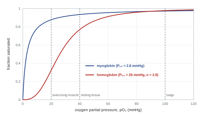
The sigmoid curve is exactly what a transport protein needs. In the lungs, where the oxygen pressure is about 100 mmHg, hemoglobin is nearly saturated. In active tissues, where it may fall to 20 mmHg, hemoglobin's saturation drops steeply and it releases much of its oxygen. A protein with myoglobin's high affinity would still be almost full at 20 mmHg and deliver very little. Hemoglobin's affinity is also tuned by the conditions in the tissues. Protons and carbon dioxide, which are abundant in active muscle, stabilize the T state and lower its affinity, an effect named after Christian Bohr, who described it in 1904. The molecule 2,3-bisphosphoglycerate, made in red blood cells, binds in the central cavity of deoxyhemoglobin and also lowers its affinity; its level rises within days of moving to high altitude, helping oxygen delivery. Fetal hemoglobin binds 2,3-bisphosphoglycerate weakly, so it has a higher affinity than the mother's hemoglobin and can draw oxygen across the placenta.
Carbon monoxide shows the other side of the story. It binds the heme iron about 200 times more tightly than oxygen and also locks the remaining subunits in the high-affinity state, so they hold on to their oxygen instead of releasing it in the tissues. Even a small fraction of carboxyhemoglobin therefore cuts oxygen delivery more than the loss of carrying capacity alone would suggest.
A worked example: oxygen delivery
Worked problem
How much oxygen does hemoglobin deliver?
Using the Hill equation with P₅₀ = 26 mmHg and n = 2.8, find the fractional saturation of hemoglobin in the lungs (pO₂ = 100 mmHg) and in exercising muscle (pO₂ = 20 mmHg). Compare the fraction delivered with that of a protein binding like myoglobin (P₅₀ = 2.8 mmHg, n = 1).
Show solution
- Lungs: (100/26)^2.8 = 3.846^2.8. The log of 3.846 is 0.585; times 2.8 gives 1.638, so the ratio is 10^1.638 = 43.5. Y = 43.5/(43.5 + 1) = 0.978.
- Muscle: (20/26)^2.8 = 0.769^2.8. The log of 0.769 is −0.114; times 2.8 gives −0.319, so the ratio is 0.480. Y = 0.480/1.480 = 0.324.
- Hemoglobin delivers 0.978 − 0.324 = 0.654, about 65% of its maximum load, between the lungs and the muscle.
- For a myoglobin-like protein: Y in the lungs = 100/102.8 = 0.973; in the muscle Y = 20/22.8 = 0.877. It would deliver only 0.973 − 0.877 = 0.096, about 10%.
Answer: Hemoglobin falls from about 98% to 32% saturated and releases about 65% of its oxygen; a non-cooperative, high-affinity carrier would release under 10%.
A program: cooperative binding
The program tabulates the saturation of myoglobin and hemoglobin over a range of oxygen pressures, then calculates how much oxygen each would deliver between the lungs and resting or exercising tissue. It also shows the Bohr effect by raising hemoglobin's P₅₀ to 31 mmHg, as happens when the pH of active muscle falls.
def hill(p, p50, n):
    return p ** n / (p ** n + p50 ** n)

MB = (2.8, 1.0)       # myoglobin: P50 (mmHg), Hill coefficient
HB = (26.0, 2.8)      # hemoglobin at pH 7.4
HB_ACID = (31.0, 2.8) # hemoglobin in active muscle (Bohr effect)

print("pO2 (mmHg)   myoglobin   hemoglobin")
for p in (5, 10, 20, 26, 40, 60, 100):
    print(f"{p:10d} {hill(p, *MB):11.3f} {hill(p, *HB):12.3f}")

print()
lungs = 100
for name, tissue in (("resting tissue", 40), ("exercising muscle", 20)):
    for label, params in (("myoglobin-like", MB), ("hemoglobin", HB), ("hemoglobin, Bohr shift", HB_ACID)):
        delivered = hill(lungs, *params) - hill(tissue, *params)
print(f"{name:18s} {label:23s} delivers {delivered:5.1%} of capacity")pO2 (mmHg) myoglobin hemoglobin
5 0.641 0.010
10 0.781 0.064
20 0.877 0.324
26 0.903 0.500
40 0.935 0.770
60 0.955 0.912
100 0.973 0.978
resting tissue myoglobin-like delivers 3.8% of capacity
resting tissue hemoglobin delivers 20.8% of capacity
resting tissue hemoglobin, Bohr shift delivers 29.2% of capacity
exercising muscle myoglobin-like delivers 9.6% of capacity
exercising muscle hemoglobin delivers 65.3% of capacity
exercising muscle hemoglobin, Bohr shift delivers 73.7% of capacityAt rest, with tissue oxygen at 40 mmHg, hemoglobin delivers about a fifth of its load and keeps the rest as a reserve. In exercising muscle it delivers about two thirds, and the Bohr shift adds roughly another eight points, freeing more oxygen exactly where acid is being made. The myoglobin-like carrier barely unloads at all. Myoglobin's high affinity is right for its own job, which is to accept oxygen from hemoglobin and hold it in the muscle cell until the oxygen pressure falls very low during intense work.
Common mistakes
MistakeSecondary structure is held together by side-chain interactions.
InsteadAlpha helices and beta sheets are held by hydrogen bonds between backbone C=O and N–H groups; side chains decide which structure forms.
MistakeA denatured protein has had its peptide bonds broken.
InsteadDenaturation destroys the fold, not the sequence; peptide bonds remain intact, and some proteins can refold.
MistakeFolded proteins are extremely stable structures.
InsteadNet stability is small, typically 20 to 60 kJ/mol, because many weak interactions are offset by the entropy lost on folding.
MistakeHemoglobin is just four myoglobins packed together.
InsteadIts subunits interact, so binding at one site changes the others; that cooperativity gives the sigmoid curve that myoglobin lacks.
MistakeA higher oxygen affinity always makes a better oxygen carrier.
InsteadA carrier must also release oxygen; too high an affinity means little is delivered to the tissues.
MistakeProteins fold by trying every possible shape until they find the right one.
InsteadA random search would take too long; folding follows an energy funnel, often helped by chaperones.
Summary and key terms
- Protein structure is described at four levels: sequence, local backbone structure, the fold of one chain, and the assembly of several chains.
- Alpha helices and beta sheets are held by backbone hydrogen bonds; proline and glycine disrupt helices.
- The hydrophobic effect drives folding, and hydrogen bonds, salt bridges, van der Waals contacts and disulfides stabilize the fold, which is only marginally stable.
- The sequence specifies the fold (Anfinsen); folding follows an energy funnel, assisted in cells by chaperones, and misfolding can cause disease.
- Hemoglobin binds oxygen cooperatively, giving a sigmoid curve tuned by protons, CO₂ and 2,3-bisphosphoglycerate for efficient delivery.
- Alpha helix
- A right-handed coil of backbone with 3.6 residues per turn, held by hydrogen bonds from each C=O to the N–H four residues along.
- Beta sheet
- Extended strands side by side, linked by backbone hydrogen bonds, running parallel or antiparallel.
- Domain
- A part of a protein chain that folds independently into a compact unit.
- Denaturation
- Loss of the folded structure of a protein without breaking its peptide bonds.
- Chaperone
- A protein that helps other proteins fold correctly and prevents them from clumping.
- Cooperative binding
- Binding in which one ligand bound makes further binding easier, giving a sigmoid curve.
- Bohr effect
- The lowering of hemoglobin's oxygen affinity by protons and carbon dioxide.
Practice problems
Work each problem on paper first, then reveal the answer to check.
How long is an alpha helix of 36 residues?
Show answer
36 × 0.15 nm = 5.4 nm, or 10 turns.
Why is proline rare in the middle of alpha helices?
Show answer
Its nitrogen has no hydrogen to donate a hydrogen bond, and its ring kinks the backbone.
What fraction of myoglobin is saturated at 10 mmHg?
Show answer
Y = 10/(10 + 2.8) = 0.78.
Explain why adding urea to a protein solution can unfold it while leaving its sequence unchanged.
Show answer
Urea disrupts hydrogen bonds and makes buried hydrophobic groups less unfavourable in water, so the weak interactions holding the fold give way.
In sickle-cell hemoglobin, which residue replaces glutamate at position 6 of the beta chain, and why does it matter?
Show answer
Valine; the hydrophobic valine creates a sticky patch that lets deoxygenated hemoglobin polymerize into fibres.
Which state of hemoglobin, T or R, has the higher oxygen affinity?
Show answer
The R (relaxed) state.
Lesson quiz · 5 questions
Score 60% or more to complete this lesson.
Create a free account to save your progress, take quizzes and earn a certificate.