
HSE-394 · Lesson 10 of 12 · 21 min
Radiation in imaging and nuclear medicine
▶ Video coming soon A video version of this lesson is on the way.
The principles from Lesson 9 apply everywhere, but each area of the hospital that uses radiation has its own patterns of exposure and its own controls. This lesson walks through the main ones: general X-ray rooms, portable X-rays on the wards, fluoroscopy in the operating room and interventional suites, CT, nuclear medicine, and patients who carry radioactive material back to the units. For each, the key questions are the same: where does the radiation come from, who is near it, and how do time, distance and shielding apply.
The main source: scatter from the patient
In X-ray imaging, the worker is almost never in the main beam. The radiation that reaches staff is scatter: X-rays that hit the patient's body and bounce off in all directions. The patient becomes the source. Scatter is much weaker than the main beam, but it is spread around the room, and staff who stand near the patient during many procedures can accumulate a significant dose over time. Three facts follow:
- The more radiation goes into the patient, the more scatter there is. Lowering the patient dose, with good technique, short beam-on times and the smallest field size, lowers staff dose too.
- Scatter is strongest on the side of the patient where the beam enters, which in most fluoroscopy is the side nearest the X-ray tube.
- Scatter follows the inverse square law from the patient, so stepping back works.
General X-ray rooms
Fixed X-ray rooms are designed with shielded walls, doors and a control area behind a leaded window, so that the medical radiation technologist stands behind shielding during every exposure. Ontario's X-ray Safety regulation and the Healing Arts Radiation Protection Act require X-ray equipment to be registered and installed according to an approved plan, with shielding checked by a qualified expert. Lights and signs at the door warn when the beam is on, and nobody else should be in the room during an exposure unless they are holding or helping the patient and wearing protection.
Portable X-rays on the units
Portable X-ray machines bring imaging to patients who cannot be moved: in intensive care, the emergency department, operating rooms and wards like 4 West. Here, nurses, other patients and visitors can be close by. The doses are small for any one exposure, but the habits matter because the exposures add up.
- Before the exposure, the medical radiation technologist calls out a warning, such as "X-ray", so others can step away.
- Staff who do not need to be there move at least 2 m (6 ft) from the patient, out of the direct line of the beam. At that distance the scattered dose from one portable exposure is tiny.
- Staff who must stay close, for example to hold a patient's position, wear a lead apron and stand to the side, not in line with the beam.
- Other patients in a shared room are usually at least 2 m (6 ft) away; curtains give privacy but no shielding.
- Pregnant workers step out of the room.
At Northbridge, nurses on 4 West used to leave the room for every portable X-ray, even when it meant leaving an unstable patient alone. After Dr. Ward showed them the dose arithmetic from Lesson 9, the unit changed its practice: nurses step back to the foot of the bed or the doorway, about 2 to 3 m (6 to 10 ft) away, and stay in sight of the patient. Their doses, measured with badges for three months, stayed at background.
Fluoroscopy
Fluoroscopy uses a continuous or pulsed X-ray beam to produce live moving images. It is used in interventional radiology, cardiac catheterization labs, the operating room (for orthopedic and vascular surgery, often with a C-arm), endoscopy and pain clinics. Procedures can last an hour or more, with staff standing beside the patient, which makes fluoroscopy the main source of occupational radiation dose in most hospitals.
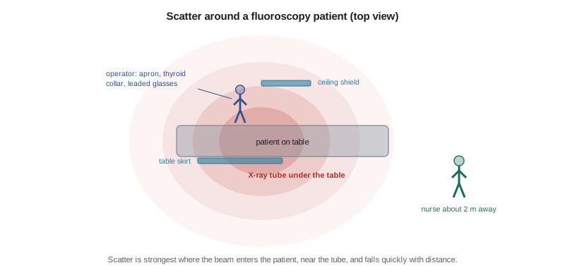
Controls in fluoroscopy work at several levels:
- Equipment and technique: pulsed fluoroscopy at the lowest pulse rate that gives an adequate image, last-image hold instead of more beam time, tight collimation to the area of interest, keeping the image receptor close to the patient and the tube as far away as practical, and the tube under the table so the strongest scatter goes toward the floor.
- Fixed and mobile shielding: table-mounted lead skirts that block scatter from below, ceiling-suspended leaded acrylic shields positioned between the patient and the operator's head, and mobile shields for staff who do not need to be at the table.
- Personal protection: lead or lead-equivalent aprons that wrap around the body, thyroid collars, and leaded glasses with side shields, since cataracts are the deterministic effect most often found in interventional staff. Aprons are checked for cracks every year and stored hanging, not folded.
- Distance and time: staff who are not needed at the table stand back; the operator keeps hands out of the beam; beam-on time is recorded and reviewed.
- Monitoring: collar badges outside the apron, with ring badges or electronic dosimeters for operators.
CT
CT scanners deliver higher patient doses than plain X-rays, but staff are rarely exposed, because the scan is run from a shielded control room. The exceptions are CT-guided procedures, where a physician may work beside the patient during scanning; the same apron, shield and distance rules as fluoroscopy apply. Family members who stay with a child during a scan wear an apron.
Nuclear medicine
Nuclear medicine uses radioactive drugs, called radiopharmaceuticals, given by injection, by mouth or by inhalation. The drug goes to an organ or a tumour, and a camera detects the gamma rays or, in PET scanning, the radiation from positrons. Some radiopharmaceuticals are used for treatment, delivering radiation directly to a cancer.
Unlike X-rays, these materials keep emitting radiation until they decay, and they can spread as contamination. Workers are exposed in two ways: external dose from vials, syringes and patients, and contamination of skin or surfaces from spills and from patients' body fluids. Common radiopharmaceuticals decay at very different rates.
| Radionuclide | Half-life | Main use | Worker concerns |
|---|---|---|---|
| Technetium-99m | About 6 hours | Most diagnostic scans (bone, heart, lung) | Dose to hands when preparing and injecting |
| Fluorine-18 | About 110 minutes | PET scans | Higher-energy emissions; shielded syringes and waiting rooms |
| Iodine-131 | About 8 days | Treatment of thyroid cancer and overactive thyroid | Patients emit radiation and excrete iodine in urine, sweat and saliva for days |
| Lutetium-177 | About 6.7 days | Treatment of some neuroendocrine and prostate cancers | Patient care, contamination and waste |
| Iodine-125 seeds | About 60 days | Prostate implants and seed localization in breast surgery | Tracking every seed; low-energy emissions |
Controls in the nuclear medicine department include syringe and vial shields made of lead or tungsten, tongs for handling vials, shielded workstations with leaded glass, drip trays and absorbent pads, disposable gloves changed often, personal and ring dosimeters, a ban on eating and drinking, and routine surveys of hands and work surfaces with a contamination meter. Radioactive waste is stored behind shielding until it has decayed, usually ten half-lives, after which it can be disposed of as ordinary waste once a survey confirms it is at background.
Decay arithmetic is simple and is used every day: after each half-life, half the radioactivity remains. A short calculation shows why technetium waste can be cleared in a few days while iodine-131 waste must be held for months.
# Radioactive decay: fraction remaining after time t
half_lives = {"Tc-99m": 6.0, "F-18": 1.83, "I-131": 8.0 * 24, "Lu-177": 6.7 * 24}  # hours

def remaining(t_hours, half_life_hours):
    return 0.5 ** (t_hours / half_life_hours)

print(f"{'nuclide':8}{'1 day':>10}{'1 week':>10}{'10 half-lives':>16}")
for name, h in half_lives.items():
    day = remaining(24, h)
    week = remaining(24 * 7, h)
    ten = 10 * h
    span = f"{ten / 24:.1f} days" if ten >= 24 else f"{ten:.0f} hours"
    print(f"{name:8}{day:10.4f}{week:10.4f}{span:>16}")
print(f"after 10 half-lives, fraction left = {0.5 ** 10:.5f} (about 1 in 1000)")nuclide 1 day 1 week 10 half-lives Tc-99m 0.0625 0.0000 2.5 days F-18 0.0001 0.0000 18 hours I-131 0.9170 0.5453 80.0 days Lu-177 0.9017 0.4847 67.0 days after 10 half-lives, fraction left = 0.00098 (about 1 in 1000)
Patients who carry radioactivity
Some of the highest nuclear medicine exposures happen outside the department, when patients return to a unit or stay in hospital after treatment. For diagnostic scans with technetium-99m, the patient's radioactivity is small and decays within a day; normal care with standard precautions is enough, though pregnant staff may limit close contact for the rest of the day.
Patients treated with large amounts of iodine-131 or lutetium-177 need more planning. Depending on the amount given and the rules that apply, the patient may stay in a shielded single room with its own bathroom, with the floor and surfaces covered, and with signs on the door. Nursing care is planned to limit time at the bedside, staff stay at a distance when they can, and care is shared among staff rather than given by one nurse. Body fluids are treated as contaminated; linen and waste are held and surveyed by the radiation safety staff before they leave the room. Pregnant staff and visitors do not enter. The patient is discharged when their measured dose rate falls below the release limit, with written instructions for home.
Patients who have had radioactive seeds implanted, such as iodine-125 seeds for prostate cancer, emit low levels of radiation that are mostly absorbed in their own bodies. Staff need no special precautions beyond knowing that a lost seed, found in a dressing or a urinal, must be picked up with forceps, placed in a shielded container and reported to the radiation safety officer, never thrown in the garbage.
In Canada, nuclear medicine departments hold a licence from the CNSC, which sets the conditions for handling, storing and disposing of radioactive material, for designating NEWs, and for releasing patients. Each licence names a radiation safety officer who runs the radiation protection program.
Spills and emergencies
A spill of a radiopharmaceutical is handled like a chemical spill with extra steps: warn others, cover the spill with absorbent material, keep people who may be contaminated in the area until they are checked, call the radiation safety officer, and do not clean up a large spill without the radiation safety staff. Contaminated skin is washed with lukewarm water and mild soap, not scrubbed. The area is surveyed after cleaning to confirm contamination is gone.
Worked problem
An iodine-131 patient on 4 West
A patient who received a treatment dose of iodine-131 in nuclear medicine this morning is admitted to 4 West overnight because of vomiting. The nurses are worried about their exposure. Rachel Kim, who is pregnant, is assigned to the patient's room. What should be done?
Show solution
- Call the radiation safety officer, Dr. Ward, before the patient arrives, so that a suitable room can be prepared: a single room with its own bathroom, covered floor and surfaces, a shielded waste container and a sign on the door.
- Reassign Rachel. Pregnant staff should not care for patients who have received therapy doses of radioactive iodine, and she should not need to explain why.
- Plan care to reduce time at the bedside and share it among staff; keep distance when possible, for example talking with the patient from the doorway.
- Treat vomit, urine and linen as radioactively contaminated: gloves, gown and shoe covers, absorbent pads, and all waste and linen held in the room for survey.
- Issue electronic dosimeters to the nurses caring for the patient, and record their doses.
Answer: Prepare a suitable room with the radiation safety officer, reassign the pregnant nurse, limit and share bedside time, handle body fluids as contaminated, and monitor the staff involved.
Common mistakes
MistakeLeaving an unstable patient alone during a portable X-ray.
InsteadStep back at least 2 m (6 ft) and stay in sight of the patient.
MistakePositioning the fluoroscopy tube above the patient without need.
InsteadKeep the tube under the table so the strongest scatter goes toward the floor.
MistakeFolding lead aprons for storage.
InsteadHang them, and have them checked for cracks every year.
MistakeThrowing a lost radioactive seed in the garbage.
InsteadPick it up with forceps, put it in a shielded container and call the radiation safety officer.
MistakeCleaning up a large radioactive spill without radiation safety staff.
InsteadCover it, keep people in place for checks and call the radiation safety officer.
Summary and key terms
- In X-ray imaging, staff are exposed mainly to scatter from the patient, strongest on the beam-entry side.
- Fixed X-ray rooms have shielded walls and control areas; portable X-rays call for staff to step back at least 2 m (6 ft).
- Fluoroscopy is the main source of staff dose; use good technique, the tube under the table, shields, aprons, thyroid collars and leaded glasses.
- Nuclear medicine materials keep emitting until they decay and can spread as contamination.
- Radioactive waste is held for about ten half-lives and surveyed before disposal.
- Patients given therapy doses need planned rooms, shared and time-limited care, and handling of body fluids as contaminated.
- Lost seeds and spills are reported to the radiation safety officer.
- Scatter
- X-rays that bounce off the patient in many directions, the main source of staff dose.
- Fluoroscopy
- Live X-ray imaging using a continuous or pulsed beam.
- C-arm
- A mobile fluoroscopy unit with the tube and receptor on a C-shaped arm.
- Radiopharmaceutical
- A radioactive drug used for diagnosis or treatment.
- Half-life
- The time for half of a radioactive material to decay.
- Contamination
- Radioactive material where it is not wanted, such as on skin or surfaces.
- Radiation safety officer
- The person named on a radiation licence who runs the radiation protection program.
Practice problems
Work each problem on paper first, then reveal the answer to check.
Where is scatter strongest during fluoroscopy?
Show answer
On the side of the patient where the beam enters, usually nearest the X-ray tube.
How far should staff step back during a portable X-ray?
Show answer
At least 2 m (6 ft), out of line with the beam.
Why are leaded glasses important for interventional staff?
Show answer
Cataracts of the lens are a common deterministic effect from years of scatter to the eyes.
How much of a radionuclide is left after ten half-lives?
Show answer
About 1 in 1000.
Why must iodine-131 waste be held much longer than technetium-99m waste?
Show answer
Iodine-131 has a half-life of about 8 days, against about 6 hours for technetium-99m.
What should a nurse do with a radioactive seed found in a dressing?
Show answer
Pick it up with forceps, place it in a shielded container and report it to the radiation safety officer.
Lesson quiz · 5 questions
Score 60% or more to complete this lesson.
Create a free account to save your progress, take quizzes and earn a certificate.